 StainList
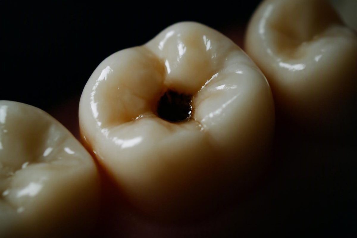
The short answer: what a small cavity may look like
A small or early area of tooth decay may look like:
- A localized dull, matte, milky, opaque, or chalky-white patch
- A persistent yellow, tan, brown, gray, or black mark
- A rough or irregular-looking area
- A tiny dent, pit, or visible opening
- Nothing visible at all, particularly when decay is between teeth or beneath an apparently intact surface
The earliest visible change may be white rather than dark. A chalky-white spot can represent demineralization—the loss of minerals from enamel—before the tooth develops a true hole. At this stage, the surface may remain intact, and there may be no pain, sensitivity, or rough texture. Cleveland Clinic distinguishes this early mineral-loss stage from the later formation of an opening in the tooth.
As damage progresses, the affected area may become discolored, rough, pitted, or visibly open. But there is no single “cavity color.” White, brown, gray, and black marks can also be stains or other enamel changes, while genuine decay can remain hidden.
Color, visible size, and appearance alone cannot confirm tooth decay. These descriptions are recognition clues, not a visual diagnostic test or a substitute for advice from a dentist. A new or changing tooth spot is a reason to arrange an assessment—not proof that you do or do not have a cavity.
How the appearance can change as tooth decay progresses
It is more accurate to think of tooth decay as a process than to treat every mark as an established cavity. In this guide, early decay refers to mineral loss that may still have an intact enamel surface. A formed cavity means that tooth structure has broken down and an opening has developed.
Plaque bacteria produce acids after exposure to sugars and starches. Repeated acid attacks remove minerals from enamel; continued damage can eventually create an opening and extend into deeper tooth layers. Early decay may be symptom-free. Harvard Health describes this progression from mineral loss to a formed cavity.
Stage one: intact white-spot mineral loss
An early area may appear:
- Duller than the surrounding enamel
- Matte rather than glossy
- Chalky, milky, or opaque white
- Localized to one part of the tooth
There may be no hole, dent, roughness, pain, or sensitivity. A white spot is not automatically decay, however. A dental examination is needed to determine what caused the color change and whether the area represents active mineral loss, a stable change, or something else.
Stage two: discoloration without an obvious hole
A suspicious area may appear yellowish, tan, light brown, gray, dark brown, or black. It may remain flat, or its surface may begin to look irregular.
Color does not reliably reveal how deep, active, or serious a lesion is. A dark mark may be staining in a natural groove, an inactive lesion, or active decay. Conversely, meaningful decay may not look particularly dark.
Stage three: visible structural breakdown
As enamel loses its integrity, the tooth may develop:
- A rough or irregular patch
- A small dent
- A pit that looks different from the tooth’s normal anatomy
- A tiny opening or more obvious hole
- An edge where food repeatedly catches
A visible opening is more concerning than discoloration alone because it suggests physical loss of tooth structure. Even then, looking in a mirror cannot establish how far the damage extends beneath the surface.
| Possible appearance | Surface condition | Possible sensations | What it can and cannot establish |
|---|---|---|---|
| Dull, chalky, or opaque white spot | Often intact and sometimes smooth | Commonly none | Can suggest mineral loss; cannot prove active decay or establish the required treatment |
| Yellow, tan, brown, gray, or black mark | May be smooth, irregular, or difficult to assess | None or occasional sensitivity | Can justify examination; color cannot determine depth or activity |
| Rough-looking or localized pitted area | Possible structural breakdown | Food trapping or sensitivity may occur | More concerning for decay, but natural anatomy and enamel changes can look similar |
| Tiny opening or visible hole | Tooth structure appears to be missing | May be painless or sensitive | Suggests cavitation but does not reveal the full extent of damage |
| No visible change | Surface may look intact | None, sensitivity, or discomfort | Does not exclude decay between teeth or below the visible surface |
There is no dependable home timeline for moving from one appearance to another.
What small cavities can look like in different parts of a tooth
Location changes both the possible appearance of decay and how easily it can be seen. A small lesion on a front-facing smooth surface may be noticeable in a mirror, while one between back teeth may remain invisible.
Smooth surfaces and the gumline
On the smooth side of a tooth or near the gumline, early mineral loss may look like a localized chalky, milky-white, or unusually matte patch. It may stand out because it looks different from the surrounding enamel.
A discolored gumline area should not automatically be labeled a cavity. Deposits, staining, exposed root structure, and non-cavity enamel changes can affect the appearance of this area. The location makes a focused dental examination worthwhile, but it does not supply a diagnosis.
Molar pits and fissures
On a molar’s chewing surface, suspicious decay may resemble:
- A persistent brown or black dot
- A dark line within a groove
- A groove that looks wider or more irregular than nearby grooves
- A small rough or pitted defect
- An area where food repeatedly catches
This is also one of the easiest places to mistake normal anatomy or staining for decay. Molars naturally have pits and fissures that can retain material and pigment. Back teeth are difficult to inspect and their grooves are harder to clean thoroughly, so a dark groove is a reason for professional checking—not a diagnosis. Mayo Clinic identifies pit-and-fissure, smooth-surface, and root cavities as distinct cavity locations.
The side of a tooth
A lesion on the visible side of a tooth may appear as a concentrated gray, brown, or black area. If the enamel has broken down, it may eventually resemble a small indentation or opening.
A mark that remains after normal brushing deserves attention, particularly if it is new or changing. Persistence does not prove decay, however, because some stains and other forms of discoloration will not brush away either.
Between teeth
Decay between teeth may occasionally create a faint shadow, gray area, or darker wedge visible through the side of a tooth. More often, it cannot be seen in a household mirror at all. The contact point where two teeth meet can conceal the affected surface while the visible outer enamel continues to look intact.
This is one reason dentists may use X-rays when hidden decay is a concern. Clean-looking spaces or a normal phone photograph cannot establish that the areas between the teeth are decay-free.
Exposed root surfaces
Cavities are not limited to enamel or chewing surfaces. They can also occur on an exposed root surface, usually close to the gumline.
Clinician-reviewed educational images can help illustrate these differences, but photographs cannot diagnose a specific tooth. When images are used, they should be clearly labeled by location and should distinguish an intact white-spot lesion, a stained groove, a cavitated lesion, root-surface decay, and between-teeth decay shown on an X-ray.
Cavity or tooth stain? Clues that help—and their limits
Cavities and stains can both be white, brown, gray, or black. Color alone cannot separate them. Texture, distribution, structural change, location, symptoms, and change over time may make decay more concerning, but none provides a definitive home test.
| Feature | Pattern that may fit staining | Pattern more concerning for decay | Important limitation |
|---|---|---|---|
| Distribution | Broad, flat discoloration across part of a tooth or several teeth | A concentrated mark or defect in one vulnerable area | Stains can be localized, and decay can affect more than one tooth |
| Texture | Appears smooth and level with nearby enamel | Looks rough, irregular, dented, or pitted | Texture is difficult to assess reliably at home |
| Structural change | No visible loss of tooth structure | A new pit, broken edge, or opening | Natural grooves and non-cavity defects can mimic damage |
| Persistence | May remain despite brushing | Also remains despite brushing | Failure to brush away is not proof of decay |
| Change over time | Long-stable and flat | New, enlarging, deepening, or changing | A stable mark may still be an inactive lesion or another change needing assessment |
| Location | May be widespread or present on exposed surfaces | Concentrated in a molar groove, near the gumline, or between teeth | Both staining and decay can occur in these locations |
| Symptoms | Often no associated sensation | Sensitivity, biting discomfort, food trapping, or pain may occur | Early cavities can be painless, and these symptoms have other causes |
A localized mark that is changing, rough-looking, pitted, or accompanied by visible structural damage is more concerning than broad, flat discoloration. A smooth or long-stable mark may be staining, an inactive lesion, or another enamel change; it should not automatically be labeled harmless. Dental-practice guidance on comparing cavities with stains likewise emphasizes that they can look nearly identical without professional assessment.
Dentists assess the tooth under controlled conditions and interpret the surface together with its location, appearance, symptoms, history, and any necessary imaging.
Natural pits and grooves can look dark, and enamel can have non-cavity variations in color or shape. Trying to diagnose every white patch or dark dot at home creates two risks: treating harmless anatomy as disease or dismissing genuine decay as a stain.
Why a small cavity may not hurt—or be visible
A cavity does not have to hurt. Early mineral loss and small lesions confined to enamel commonly cause no pain or sensitivity. A tooth can look and feel normal while decay is beginning.
Symptoms may develop as the affected area enlarges or reaches deeper tooth layers. Possible signs include:
- Sensitivity to sweet, hot, or cold foods and drinks
- Discomfort or pain when biting
- Repeated food trapping in the same place
- Toothache
- Persistent bad breath
- An unpleasant or unusual taste
These symptoms cannot diagnose a cavity. Sensitivity, biting discomfort, bad breath, food trapping, and an unusual taste all have other possible causes.
Hidden location is another problem. Decay can form at the contact point between two teeth, beneath an apparently intact surface, or inside a back-tooth groove that is difficult to see. Some lesions are found only during a dental examination or through imaging. Cleveland Clinic notes that cavities can occur on the crown or root and may not be felt until deeper layers are affected.
Do not rely on any of the following as reassurance:
- “The spot is too tiny to matter.”
- “It doesn’t hurt.”
- “I can’t see a hole.”
- “My tongue doesn’t feel anything unusual.”
- “It looks normal in a phone photo.”
A painless tooth is not necessarily decay-free, and a visually dramatic mark is not necessarily active decay. Appearance and symptoms provide useful context, but they do not replace diagnosis.
How a dentist confirms whether a spot is tooth decay
A dentist evaluates much more than color. Depending on the tooth and suspected location, assessment may involve:
- Visual inspection under controlled lighting
- Drying and examining the tooth surface
- Checking whether the enamel is intact or structurally damaged
- Using appropriate clinical instruments
- Reviewing symptoms and dental history
- Taking dental X-rays when hidden decay is a concern
The dentist considers where the mark is located, whether tooth structure has broken down, whether the appearance fits that location, whether the area has changed, and whether imaging supports decay. This combined assessment helps distinguish active disease from staining, normal anatomy, an inactive lesion, or another enamel change.
X-rays are particularly useful for examining areas between teeth or beneath a surface that appears intact. Decay generally appears darker than healthy tooth structure on an X-ray because the affected area has lower mineral density. A clinical overview of early cavity appearance and radiographic detection illustrates why a lesion may be more apparent on imaging than in a mirror.
Dentists decide when imaging is appropriate and interpret it together with the clinical examination. A reader should not try to prescribe a particular X-ray type or schedule based only on a tooth’s appearance.
Home checks have narrower value:
- A mirror may reveal a new mark but cannot show hidden depth.
- Brushing may remove debris but cannot distinguish every stain from decay.
- A tongue check may notice a substantial rough edge but cannot reliably assess enamel integrity.
- A photograph can document an obvious change but cannot establish the diagnosis.
These methods may help you explain what prompted concern. They are not substitutes for an examination.
Can an early cavity be reversed, or will it need a filling?
The answer depends largely on whether the enamel surface remains intact.
An area of early demineralization is not the same as a formed hole. When mineral loss is identified before the enamel collapses, the process may sometimes be stopped or remineralized through professionally guided preventive care, which can include fluoride and improvements in daily oral hygiene. Once an opening has formed, preventive care cannot replace the missing tooth structure. Harvard Health explains this treatment boundary between early mineral loss and a formed cavity.
This does not mean:
- Every white spot is decay
- Every early lesion can be reversed
- Toothpaste can close a visible hole
- A suspicious area should be monitored without professional input
- Every dark mark needs a filling
Once enamel has physically broken down and a true opening has formed, the missing structure generally cannot regrow. Treatment commonly involves removing decayed material and restoring the tooth, often with a filling. The appropriate approach depends on the lesion’s location, extent, activity, surface integrity, and the patient’s individual needs.
Some intact early lesions may be managed with preventive treatment and monitoring rather than an immediate restoration. That is why distinguishing discoloration, non-cavitated mineral loss, and an established hole matters. Treating every white or dark mark as a filling candidate would be inappropriate, but delaying assessment in the hope that brushing will close a visible hole is also unwise.
If a dentist recommends monitoring, ask when the area should be reassessed and what change would trigger treatment. If preventive care is recommended, follow the specific fluoride and oral-care plan provided for that tooth rather than assuming that more brushing or unsupervised products will repair structural damage.
What to do when you notice a suspicious spot
Use the appearance as a guide to your next step, not as a final diagnosis.
| What you notice | Sensible next step |
|---|---|
| Intact chalky-white patch | Arrange a dental assessment, particularly if it is new or localized. Early mineral loss may be manageable before a hole forms, but other enamel changes can also look white. |
| Persistent brown, gray, or black mark | Mention it at a dental visit; book sooner if it is new, changing, or confined to a vulnerable groove or contact area. |
| Rough or pitted-looking area | Seek timely evaluation because apparent structural change is more concerning than color alone. Do not probe it. |
| Visible hole or broken-down area | Arrange an appointment promptly. Do not expect toothpaste or brushing to replace missing tooth structure. |
| Sensitivity, biting discomfort, or repeated food trapping | Book an assessment even if no cavity is visible. These symptoms are not specific to decay but warrant investigation. |
| Toothache or mouth pain | See a dentist as soon as possible. |
| Severe throbbing pain, swelling, fever, pus, facial swelling, or a pimple-like bump on the gum | Seek prompt or urgent professional care because these may be signs of infection. |
Regular dental examinations matter even when the mouth feels fine because early decay may be painless, hidden, or easy to confuse with staining. Mayo Clinic advises seeing a dentist as soon as possible for toothache or mouth pain and notes that a beginning cavity may cause no symptoms. Its cavity guidance also explains why routine examinations remain important when the mouth feels normal.
Severe throbbing pain, fever, swelling, pus, facial swelling, or a pimple-like gum bump are not typical visual signs of a small, early cavity. They are listed separately because they may indicate advanced disease or infection requiring prompt attention. Dental guidance on cavity complications identifies severe pain, swelling, fever, and a gum bump as possible abscess warning signs.
In the meantime, continue normal oral hygiene. Do not scrape, drill, pick, bleach, or apply caustic substances to the tooth.
Frequently asked questions
Can a small cavity be white instead of brown or black?
Yes. Early enamel mineral loss may appear as a localized dull, opaque, milky, or chalky-white spot before a true opening exists. It may be painless and smooth.
White does not automatically mean decay, however, because other enamel changes can have a similar appearance. A dentist must determine whether the area represents active mineral loss, a stable change, or something else.
Is every tiny brown or black dot on a molar a cavity?
No. Molar grooves can be naturally deep or stained, and a dark dot may reflect pigment rather than active decay.
A localized mark that is enlarging, rough, pitted, sensitive, or repeatedly trapping food is more concerning, but those clues still cannot confirm a cavity. A dentist can assess whether the groove remains intact and whether examination or imaging shows decay.
Can a small cavity exist without pain or sensitivity?
Yes. Early decay commonly causes no pain or sensitivity, particularly while it is confined to enamel or hidden between teeth. Symptoms may emerge as damage enlarges, but their absence does not rule out a cavity.
Regular dental examinations are important precisely because an affected tooth may still feel normal.
What does a cavity between teeth look like?
It may occasionally create a gray shadow, subtle discoloration, or dark wedge visible through the side of a tooth. Frequently, it does not look like anything from the outside.
The contact area can hide decay from a mirror and camera, so a dentist may need an examination and X-rays to detect it.
Can fluoride heal a small cavity?
Fluoride may help remineralize an intact area of early enamel mineral loss under professional guidance. It cannot regrow tooth structure once a true hole has formed.
A formed cavity commonly needs restorative treatment, while some intact early lesions may be managed with prevention and monitoring. The deciding factor is not simply how small the spot looks, but whether the surface remains intact and what a dentist finds during assessment.
The key takeaway
A small cavity may be white, dark, rough, pitted, visibly open, or completely hidden. A painless tooth is not necessarily decay-free, and a dark spot is not automatically a cavity.
Use a new or changing tooth mark as a reason to arrange an assessment rather than an invitation to diagnose or treat the tooth at home. Only a dental professional can distinguish active decay from staining, normal anatomy, or another enamel change and determine whether preventive care, monitoring, or restoration is appropriate.